
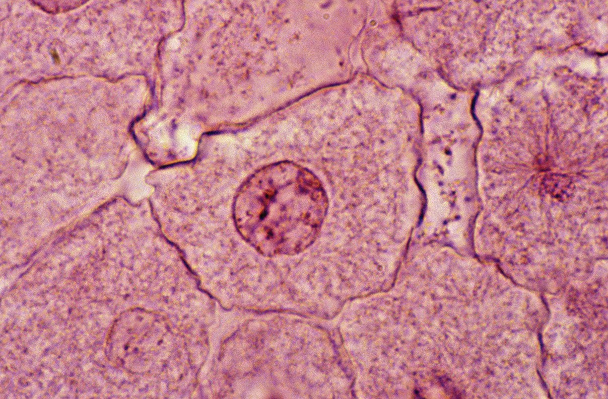
In a prokaryote, all the genes are carried in a circular DNA molecule in the cytoplasm. In a eukaryote, however, the genes are contained in a number of separate linear DNA molecules in a membrane-bound nucleus. Each of these molecules, together with associated proteins, makes a chromosome. When the nucleus is not dividing, the chromosomes are unravelled and genetically active. But when the nucleus divides at mitosis, genetic activity is switched off and the chromosomes are packaged up into highly condensed structures, which can be seen in the light microscope. The study of chromosomes is an important field of research. Preparations of condensed mitotic chromosomes, including those of humans, are easy to produce (Box 1). These have provided, and are continuing to provide, a huge amount of information about the genetic control of cell function and the mechanisms of cell division. This What is…? reviews the genetic content, composition, structure and packaging of chromosomes, and some of the consequences of variations in their number and structure, which include human disease.
A blood sample is the most convenient starting point, using white cells called T lymphocytes. These cells do not usually divide, but are stimulated to do so in liquid suspension. Only a small percentage of cells enter division, so the number is increased by the addition of agents such as colcemid. This disrupts the mitotic spindle, so preventing the separation of daughter chromosomes and holding cells in metaphase. The cells are then swollen in a hypotonic solution, chemically preserved, then burst by dropping them onto a cooled microscope slide, dried and stained. The chromosomes are then sorted into pairs according to size to produce a karyotype. The cells may also be treated to produce bands shown by stains such as Giemsa — G-banding (Figure 1). These bands are used to produce an international standard map for each chromosome known as an idiogram. Preparations can also be prepared for examination in the scanning electron microscope.
Your organisation does not have access to this article.
Sign up today to give your students the edge they need to achieve their best grades with subject expertise
Subscribe