
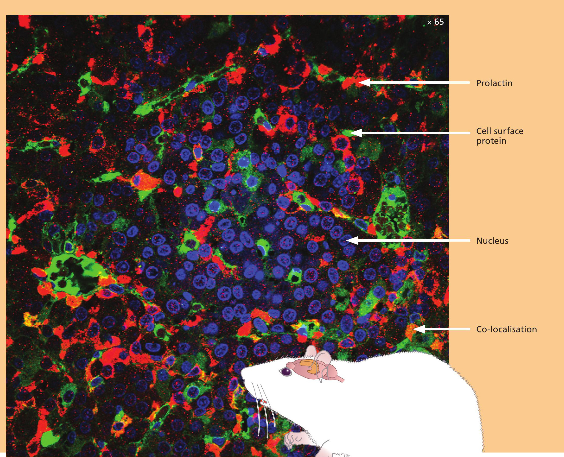
What can we tell just by looking at cells? Well actually not a lot unless we have some means of highlighting the presence of some of the cellular components. The ability to light-up parts of the cell is exactly what this image shows. Using a technique called fluorescence microscopy we can label parts of the cell with different colours (see BIOLOGICAL SCIENCES REVIEW, Vol. 24, No. 1, p. 42 for an explanation of the technique). In this instance we are looking at a cross-section of a rat pituitary gland viewed in a confocal microscope. The nucleus is labelled in blue, a cell-surface protein in green and the hormone prolactin in red. The orange colour indicates that the cell-surface protein is present on cells that are secreting prolactin, since where the two proteins co-localise, their red and green colours combine to give an orange colour.
Confocal microscopy allows us to visualise these components in specimens that are sufficiently thick to show the interior and the exterior of the cell at the same time. Using this approach scientists can learn more about where certain proteins are located and whether two proteins can be found together in/on the same cell. Information about cell morphology and where cells are located in tissues can also be obtained.
Your organisation does not have access to this article.
Sign up today to give your students the edge they need to achieve their best grades with subject expertise
Subscribe