
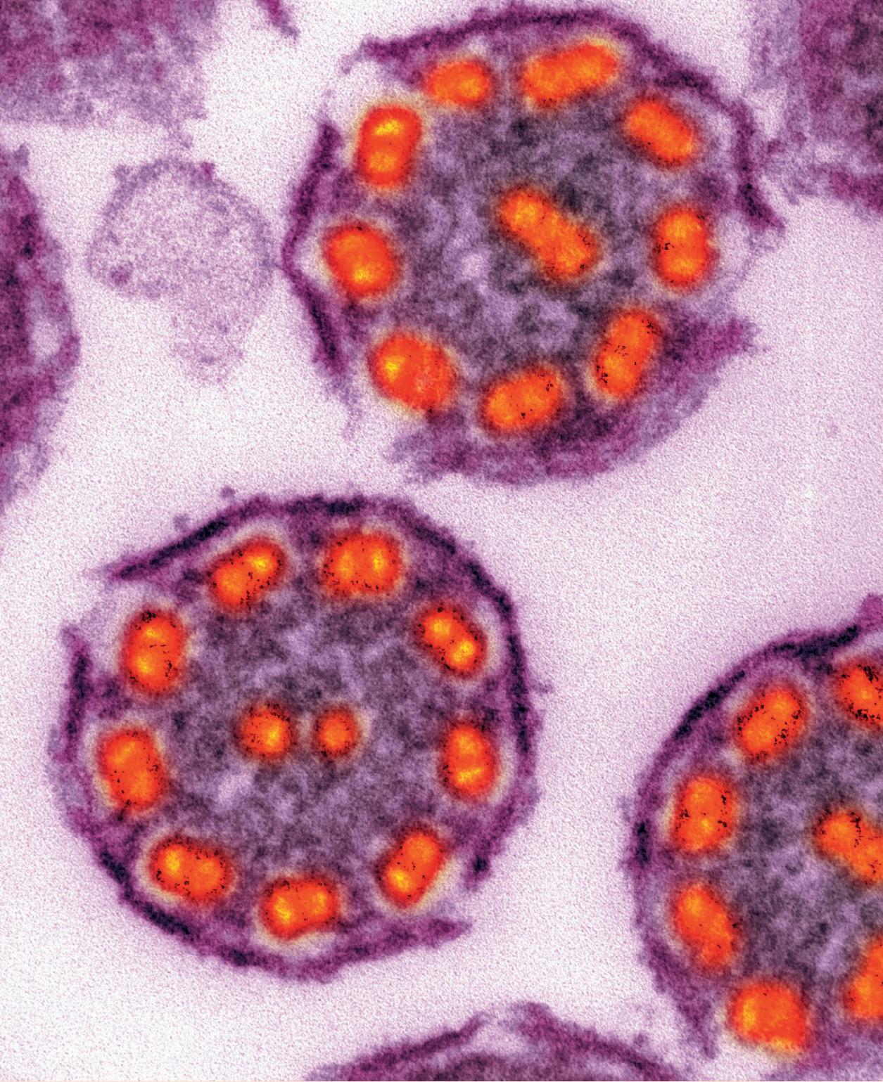
Cilia and flagella (singular: cilium and flagellum) are fine, hair-like structures found on the surface of a wide range of cells. In eukaryotic cells, the structure of cilia and flagella is similar. In cross-section they show a ‘9+2’ arrangement, comprising nine pairs of protein microtubules in a ring, with two further microtubules in the centre (see Figure 1), all enclosed by the cell-surface membrane. Movement — bending — is caused by protein arms that link adjacent pairs of microtubules and make them slide past each other. This process uses energy from the hydrolysis of ATP.
The most obvious difference between cilia and flagella is their length. Cilia are short (2–10 micrometres) and occur in large numbers, making the cell appear hairy. Flagella are longer (up to more than 1000 micrometres) and there are only a few per cell. Their most obvious function is to generate movement, but they also move differently. Cilia beat like oars, at about 5–10 beats per second, and usually in phase with each other. This leads to coordinated movement of the whole cell (see Figure 2 and http://tinyurl.com/pnodyjo) or moves fluids or particles across the cell surface (see Figures 3 and 4). Flagella move individually in an undulatory pattern, with a wave of bending passing along the length. An example is the tail of sperm (see Figure 5).
Your organisation does not have access to this article.
Sign up today to give your students the edge they need to achieve their best grades with subject expertise
Subscribe