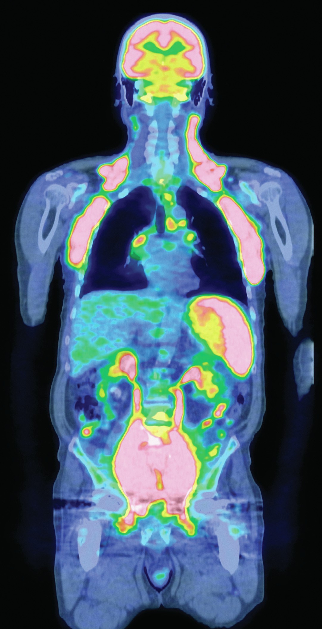
Positron emission tomography (PET) is a medical imaging technique that works through the detection of gamma radiation (Figure 1). A radiotracer agent is administered to the patient, and the emission from this can then be detected and used to provide a three-dimensional image. Through careful design of the radiotracer, different types of tissue or organs can be targeted.
18F-FDG (fluorodeoxyglucose, see Figure 2), which incorporates the 18F isotope, is a radiotracer analogue of the sugar molecule glucose. The amount of radiotracer taken up by a cell depends on its metabolic rate. Cells that have a higher metabolic rate than others, for example those found in the brain and kidneys, take up more of the tracer. Cancer cells also have an increased metabolic rate and hence greater sugar uptake, so 18F-FDG can be used to locate and visualise tumours.
Your organisation does not have access to this article.
Sign up today to give your students the edge they need to achieve their best grades with subject expertise
Subscribe